

+918048037212

Currently it only shows your basic business info. Start adding relevant business details such as description, images and products or services to gain your customers attention by using Boost 360 android app / iOS App / web portal.
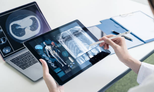
I believe you meant to refer to "ultrasonography, " which is a medical imaging technique commonly known as ultrasound. Ultrasonography uses high-frequency sound waves to create real-time images of internal body structures. It is a non-invasive and safe imaging modality widely used in various medical specialties. Here's how ultrasonography works: Transducer and sound waves: An ultrasound machine consists of a handheld device called a transducer. The transducer emits high-frequency sound waves into the body. These sound waves, typically in the range of 2 to 18 megahertz (MHz), are beyond the audible range of human hearing. Sound wave propagation: The sound waves emitted by the transducer penetrate the body tissues and bounce back (echo) when they encounter different tissues or structures with varying densities. The echoes are then picked up by the transducer. Echo detection and processing: The transducer contains receivers that detect the returning echoes. The echoes are converted into electrical signals and sent to the ultrasound machine for processing. Image formation: The ultrasound machine processes the electrical signals and constructs a real-time image based on the time it takes for the sound waves to travel and the intensity of the echoes. The machine calculates the distance and position of the reflecting structures to generate a two-dimensional (2D) or three-dimensional (3D) image. Image display and interpretation: The resulting ultrasound images are displayed on a monitor, allowing healthcare professionals, such as radiologists or sonographers, to analyze and interpret the structures and identify any abnormalities or pathologies. Ultrasonography offers several advantages: Non-invasiveness: Ultrasound imaging is non-invasive, meaning it does not involve the use of ionizing radiation or the need for surgical incisions. It is generally considered safe and painless. Real-time imaging: Ultrasound provides real-time imaging, allowing dynamic evaluation of structures such as heart valves, blood flow, fetal movement, or organ motion. Portability: Ultrasound machines are relatively portable compared to other imaging modalities, enabling their use in various healthcare settings, including clinics, hospitals, emergency rooms, and even remote or mobile locations. Versatility: Ultrasonography can examine various body structures, including the abdomen, pelvis, heart, blood vessels, thyroid, musculoskeletal system, and more. It is also commonly used for obstetric and gynecological evaluations. Cost-effectiveness: Ultrasonography is generally more cost-effective than other imaging techniques like computed tomography (CT) or magnetic resonance imaging (MRI). It can often provide valuable diagnostic information without the need for additional imaging modalities. Ultrasonography has some limitations, such as limited penetration through bone and air-filled structures, operator dependency, and challenges in obtaining high-quality images in obese or technically difficult patients. However, it remains an essential and widely used imaging modality due to its safety, versatility, real-time imaging capabilities, and wide range of applications in medical diagnostics and interventions.
